
Mycoplasmas: little bacteria with a big impact
- 5 Minutes to read
- Written by Axonlab
- General Practice
Bacteria are tiny, single-celled organisms and among the oldest forms of life. Their cell structure is very simple.
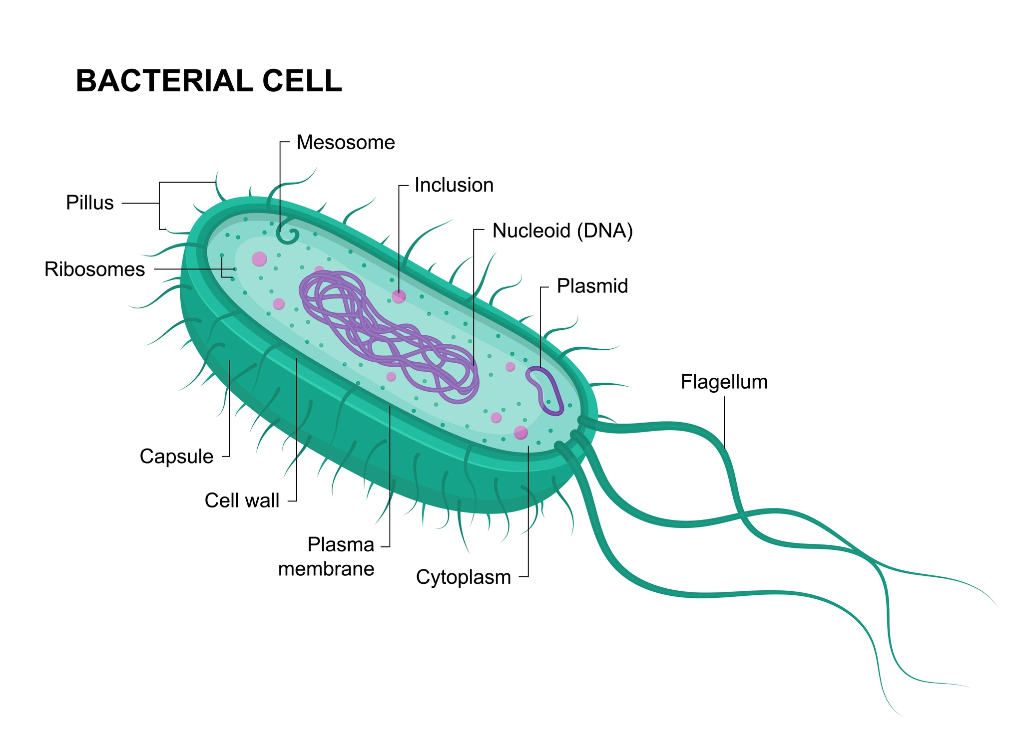
Figure 1
As you can see in Figure 1, bacteria possess a very simple structure, as they do not have a nucleus. Their genetic information is located directly inside the cell itself. Most bacteria are surrounded by a protective cell wall, which lends them their shape, and a cell membrane, which controls the flow of material in and out of the cell. Many bacteria have either a flagellum for movement or a protective layer for better defence. They are highly adaptable and can be found virtually everywhere – in our bodies too.
There are many different species of bacteria, but today we are going to be discussing mycoplasmas. Mycoplasmas are a unique type of bacteria that differ significantly from other species in the following ways:
- They do not have a cell wall, which means that not all antibiotics – including penicillin – can be used for treating them.
- They range in size between 150 and 350 nm. They are small bacteria, in other words.
- They thrive in a wide range of environments: Mycoplasmas can be found in humans, animals and plants. In humans they often colonise the respiratory tract, urogenital tract and other mucous membranes.
- They grow slowly: They grow much slower than other bacteria and can only be cultured using special media containing specific nutrients.
Figure 2 shows an image of mycoplasmas (taken with a microscope):
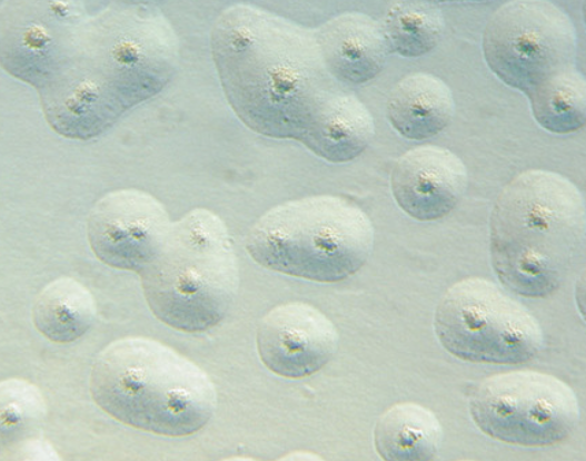
Figure 2
There are three important subgroups of mycoplasmas that can contribute to disease in healthy people:
- Mycoplasma pneumoniae
- Mycoplasma hominis
- Mycoplasma genitalium
The latter two pathogens are generally found in cases of urogenital infections.
Mycoplasma pneumoniae, by contrast, is among the five most common (bacterial) pathogens responsible for respiratory infections. This species of mycoplasmas can cause mild to moderate respiratory diseases such as bronchitis or atypical pneumonia. The latter is also referred to as “walking pneumonia.”
The bacterium adheres to the mucous membranes and attacks our natural defence mechanism: the cilia. These tiny, fine, hair-like structures are located on our cells, where their movement transports mucus, particles and pathogens out of their environments. The bacterium then releases toxic substances that weaken our cells’ defences, potentially triggering the body’s inflammatory response. The destruction of mucous membrane cells and the presence of mycoplasmas lead to an inflammatory reaction in our tissues. The immune system responds by increasing its production of proinflammatory substances that can cause symptoms such as coughing, sore throat or fever, as well as a general feeling of malaise.
The rise in mycoplasma infections in Switzerland in 2024 was the subject of numerous media reports. On 8 August 2024, for example, SRF reported an increase in infections among children – infections caused by mycoplasmas. On 19 September 2024, the Medical Tribune likewise published a report on growing case numbers of mycoplasma pneumonia in children and adults in Switzerland. Most impacted were children and teens who were in close contact with each other in schools, athletic organisations or child-care facilities – mycoplasmas are transmitted through the droplets produced by coughing, sneezing or speaking.
Sources:
- Swiss Radio and Television (SRF). (2024, August 8). Return of Mycoplasma: Record Levels of Childhood Pneumonia. Retrieved on January 20, 2025, from https://www.srf.ch/news/schweiz/rueckkehr-der-mykoplasmen-rekordwerte-bei-kinder-lungenentzuendungen
- Medical Tribune. (2024, September 19). Mycoplasma Pneumonia in Children and Adults. Retrieved on January 20, 2025, from https://medical-tribune.ch/news/medizin/4000118264/mykoplasmen-pneumonie-bei-kindern-und-erwachsenen/?
- Figure 2: Wiley Online Library. (2013). Mycoplasma Pneumonia in Children and Adults. Retrieved on January 20, 2025, from https://onlinelibrary.wiley.com/doi/abs/10.1002/biuz.201390114